Case #1
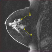
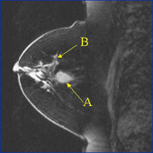
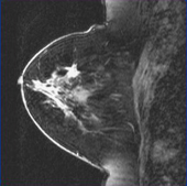
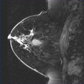
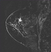
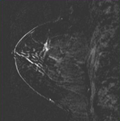
The following case shows magnetic resonance (MR) breast imaging of a patient with a benign fibroadenoma (a) and invasive lobular carcinoma (b). The two images (left and right) are taken from different planes before contrast injection (gadolinum) and then after contrast injection. The final set of pictures shows the post-contrast images subtracted from the pre-contrast images in order to display regions that are highlighted by the contrast. In particular, the subtracted images highlight the spiculated area that shows invasive lobular carcinoma. A subsequent biopsy on the patient confirmed the presence of cancer.
Before contrast injection: