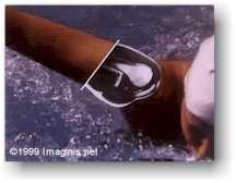
Arthroscopy is inspection through an endoscope (a flexible viewing tube) of the interior of a joint. Endoscopes now use fiber optic cables and powerful lens systems to provide improved lighting and better visualization of the interior of a joint. Endoscopes may be used in conjunction with a camera or video recorder to document images of the inside of the joint or chronicle an arthroscopic procedure. New endoscopes have digital capabilities for manipulating and enhancing images.
Arthroscopy enables orthopedic surgeons to view the surfaces of the bones that come into contact in a joint, the ligaments and cartilage within a joint, and the synovial membrane that lines the internal surface of the joint capsule. Arthroscopy can reveal diseased tissue, ligaments, and cartilage. The surgeon also can see cysts and evidence of rheumatoid and degenerative arthritis, and foreign bodies associated with gout and other disorders. Specimens of these joint structures can be removed for examination and analysis using endoscopic biopsy.